

Presented by Zia H Shah MD
Is this a new and a big domain to establish guided evolution as opposed to blind evolution, purely directed by random chance? God is not dead!
1. Introduction: The Chimeric Nature of the Vertebrate Genome
The prevailing narrative of vertebrate evolution has long been one of gradual, endogenous modification—a slow accumulation of point mutations and chromosomal rearrangements driven by natural selection acting upon the organism’s own genetic lineage. However, the advent of high-throughput genomics has radically deconstructed this view, revealing that the genomes of mammals and reptiles are not biologically hermetic seals but rather porous entities, heavily colonized by external genetic agents. Foremost among these are Endogenous Retroviruses (ERVs), the fossilized remnants of ancient germline infections that have become stable, heritable components of the host architecture.1
These viral sequences, often dismissed in early genetic theory as “junk DNA” or “genomic parasites,” constitute approximately 8-10% of the mammalian genome. While the vast majority of these elements have succumbed to mutational degradation over millions of years, becoming inert genomic fossils, a significant subset has been “co-opted” (or exapted) by the host. Through a process of “molecular domestication,” these viral genes—specifically those encoding the structural proteins Gag (capsid), Pol (polymerase), and Env (envelope)—have been repurposed to perform essential physiological functions.2
This report provides an exhaustive, expert-level analysis of the retroviruses and viral genes that have assumed positive, functional roles within the genomes of mammals and reptiles. It systematically categorizes these elements by their physiological contributions, ranging from the architectural foundation of the placenta and the complexification of the neuronal network to the fortification of the innate immune system. Furthermore, it explores the emerging comparative genomics between mammals and reptiles, highlighting the disparate yet convergent strategies employed by these lineages in harnessing the viral “enemy” for evolutionary gain.
1.1 The Mechanism of Endogenization and Co-option
Retroviruses are unique among pathogens in their requirement to integrate a DNA copy of their RNA genome (the provirus) into the host’s chromosomal DNA as an obligatory step in their replication cycle. When this integration occurs in a germ cell (sperm or egg) and the resulting embryo survives to reproduce, the retrovirus becomes “endogenous”—passed vertically to offspring as a Mendelian trait.4
The transition from a pathogenic invader to an essential symbiotic partner involves a complex evolutionary trade-off. The host must suppress the deleterious effects of the virus—such as uncontrolled replication, insertional mutagenesis, and oncogenesis—while simultaneously preserving and harnessing its useful properties.
- Env (Envelope): Originally designed to fuse the viral membrane with the host cell membrane for infection, these proteins have been co-opted to fuse host cells together (syncytium formation) and to suppress the host immune system (tolerance).5
- Gag (Group-specific antigen): Originally the structural scaffold of the viral capsid, these proteins have been repurposed to transport host RNA between cells or to form protective shells around specific nucleic acids.7
- LTRs (Long Terminal Repeats): The viral promoters and enhancers have been co-opted to drive the expression of host genes, particularly in response to immune signals like interferons.9
This phenomenon is not a singular accident but a widespread, recurrent theme in vertebrate evolution, occurring independently across diverse lineages.
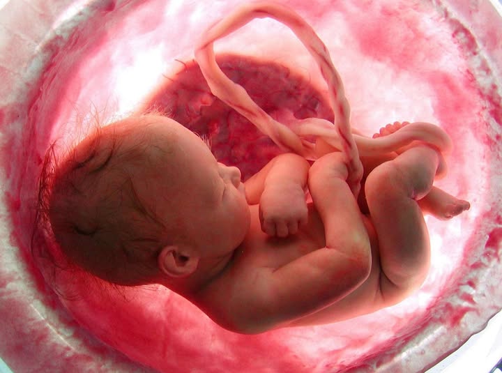
2. The Placental Revolution: Viral Envelopes as Morphogens
The evolution of the placenta in eutherian mammals and certain viviparous reptiles represents one of the most significant leaps in vertebrate history, allowing for prolonged fetal development within the maternal environment. This physiological innovation presents two formidable challenges:
- Structural Integration: The need to form a seamless interface for nutrient and gas exchange, often requiring the fusion of trophoblast cells into a multinucleated syncytium (the syncytiotrophoblast).
- Immunological Tolerance: The necessity of preventing the maternal immune system from rejecting the fetus, which is genetically semi-allogeneic (foreign).
Retroviral Envelope (Env) proteins are pre-adapted by millions of years of viral evolution to solve exactly these problems. They possess fusogenic domains to merge membranes and Immunosuppressive Domains (ISDs) to inhibit host immune responses. Consequently, the “capture” of viral env genes has driven the emergence of the placenta in multiple independent lineages.1
2.1 The Syncytin Genes: A Case Study in Convergent Evolution
The “Syncytin” family of genes is not a single genetic lineage but a functional category describing distinct viral genes captured independently by different mammalian orders. This convergence underscores the structural necessity of viral proteins for placentation.
2.1.1 Primate Syncytins: Architects of the Human Placenta
In humans and other Simiiformes (monkeys and apes), two distinct Env proteins are essential for placental development. These genes are regulated by complex interactions with other co-opted viral elements.
- Syncytin-1 (HERV-W):
- Origin: Derived from the Human Endogenous Retrovirus W (HERV-W) family, captured approximately 25 million years ago (Mya).12
- Function: It is a potent fusogen that drives the fusion of cytotrophoblast cells into the syncytiotrophoblast layer, the primary barrier and exchange surface of the human placenta.
- Mechanism: Syncytin-1 binds to the SLC1A5 (ASCT2) receptor, a neutral amino acid transporter, to trigger fusion.13
- Regulation: Its expression is enhanced by a specific RNA motif in its 3′ untranslated region (UTR) known as the Syncytin Post-transcriptional Regulatory Element (SPRE). This element, also found in other syncytins, is a viral remnant that facilitates high-level protein expression, a trait necessary for the massive tissue remodeling of the placenta.14
- Syncytin-2 (HERV-FRD):
- Origin: Derived from the HERV-FRD family, captured earlier than Syncytin-1, roughly 40 Mya.12
- Function: While also fusogenic, Syncytin-2 is highly conserved for its Immunosuppressive Domain (ISD). It is believed to be the primary agent inducing maternal immune tolerance toward the fetus, preventing rejection.12
- Clinical Relevance: Reduced expression of Syncytin-2 (and Syncytin-1) is strongly correlated with pre-eclampsia, a condition characterized by poor placentation and maternal hypertension, highlighting the gene’s ongoing necessity for reproductive health.12
2.1.2 Rodentia: The Murine and Sciurid Paradigms
The order Rodentia, specifically the family Muridae (mice and rats), utilizes a completely different set of retroviral genes, demonstrating that the “idea” of using a virus was more important than the specific virus used.
- Syncytin-A and Syncytin-B (Muridae):
- Origin: Derived from Murine Endogenous Retroviruses (MuERVs) captured ~20-25 Mya.5
- Syncytin-A: Essential for the formation of Syncytiotrophoblast Layer I (ST-I). Knockout mice for Syncytin-A die in utero due to gross placental defects and failure of the labyrinthine structure.12
- Syncytin-B: Required for the formation of Syncytiotrophoblast Layer II (ST-II). Interestingly, Syncytin-B knockout mice can survive to term but exhibit fetal growth retardation and placental abnormalities. Syncytin-B also possesses an immunosuppressive domain and has been implicated in non-placental fusion events, such as osteoclast formation (bone resorption).19
- Syncytin-Mar1 (Sciuridae):
- Origin: In the squirrel-related clade (Sciuridae), specifically identified in the woodchuck/marmot (Marmota monax), a distinct gene named Syncytin-Mar1 was captured 35-50 Mya.
- Function: It is fusogenic and expressed at the fetomaternal interface. Phylogenetic analysis confirms it is unrelated to the murine Syncytin-A/B or primate Syncytins, representing yet another independent capture event within Rodentia.21
2.1.3 Carnivora: Syncytin-Car1 and the Hyena Anomaly
The order Carnivora (dogs, cats, bears, seals) presents a unified capture event that predates the diversification of the order.
- Syncytin-Car1:
- Origin: Captured in the common ancestor of Carnivora, approximately 60-85 Mya.23
- Distribution: Found in dogs (Canis lupus familiaris), cats (Felis catus), and pandas, but notably absent in the related order Pholidota (pangolins).23
- Function: It drives the formation of the syncytium in the endotheliochorial placenta characteristic of carnivores.24
- Regulation: Like primate syncytins, Syncytin-Car1 contains an SPRE-like element in its 3′ UTR, suggesting a convergent regulatory strategy where the host maintains viral RNA processing signals to ensure efficient translation.14
- Hyena-Env2 (The Hyena Exception):
- The spotted hyena (Crocuta crocuta) possesses a highly invasive hemochorial placenta (similar to humans) rather than the standard carnivore endotheliochorial type.
- In addition to Syncytin-Car1, hyenas express a lineage-specific Hyena-Env2. This gene is non-fusogenic but expressed at the fetomaternal interface, likely contributing to the specific structural or immunological requirements of the hyena’s unique placental architecture.17
2.1.4 Ruminants, Lagomorphs, and Afrotherians
The ubiquity of syncytin capture extends to almost every major clade of eutherian mammals.
- Ruminants (Syncytin-Rum1 & Fematrin-1):
- Cows and sheep have a synepitheliochorial placenta where fusion is restricted to specific binucleate cells that migrate and fuse with maternal epithelium.
- Syncytin-Rum1: Captured ~20 Mya, this gene is found in cattle and sheep and facilitates this specific fusion event.14
- Fematrin-1: Identified in Bos taurus (cattle), this gene is derived from an endogenous JSRV-like virus (Jaagsiekte sheep retrovirus) and exhibits fusogenic activity, acting alongside or independently of Rum1.26
- Other BERVs: Bovine Endogenous Retroviruses (BERV-K1, BERV-K2) are expressed in the placenta but lack fusogenic activity, potentially serving regulatory roles or structural support.14
- Lagomorphs (Syncytin-Ory1):
- Origin: Found in the rabbit (Oryctolagus cuniculus) but absent in pikas (Ochotonidae), placing its capture between 12-30 Mya.28
- Function: A classic fusogenic Env protein essential for the rabbit’s placental development.
- Afrotheria (Syncytin-Ten1):
- Found in Tenrecs (Echinops telfairi), a primitive lineage of Afrotherian mammals. Syncytin-Ten1 confirms that viral capture occurred even in the most basal branches of the placental tree. It also contains the SPRE regulatory motif.15
2.1.5 Marsupials: The Primitive Placenta and the Opossum
For decades, it was believed that syncytins were exclusive to eutherian (placental) mammals. However, the discovery of syncytins in marsupials revolutionized this view. Marsupials possess a short-lived but functional placenta before the underdeveloped young migrate to the pouch.
- Syncytin-Opo1:
- Origin: Identified in the gray short-tailed opossum (Monodelphis domestica).
- Function: It is fusogenic and specifically expressed at the placental fetomaternal interface during the brief gestation window.32
- Implication: This suggests that the “capture” of viral fusogens was likely the founding event that allowed the transition from oviparity to viviparity in the common ancestor of Therian mammals (Marsupials + Eutherians), or at least that the strategy is so advantageous it arose immediately in both lineages.33
- Other Marsupial Envs: Additional captured Env genes, such as Syncytin-Mar1 12, Env-Aja, and Env-Psc, have been identified in wallabies and other marsupials, indicating a widespread dependence on viral genes even in “primitive” placentas.34
2.2 The Reptilian Convergence: The Mabuya Skink
Perhaps the most definitive proof of the “viral necessity” for placentation comes from the Class Reptilia. While most reptiles are oviparous (egg-laying), the Mabuya skinks (Trachylepis sp.) have evolved a complex, highly invasive chorioallantoic placenta that structurally resembles the mammalian placenta.
- Syncytin-Mab1:
- Discovery: Researchers identified a retroviral env gene, named Syncytin-Mab1, in the Mabuya genome.
- Characteristics: It is expressed specifically in the placental syncytium and exhibits fusogenic activity in cell assays, identical to mammalian syncytins.36
- Mechanism: It utilizes the MPZL1 gene as a receptor to drive fusion.37
- Significance: This discovery demonstrates that placental evolution—whether in a lizard ~25 Mya or a primate ~40 Mya—is convergently reliant on the domestication of retroviral envelope proteins. The virus is not just a tool for mammals; it is the universal “key” to viviparity in vertebrates.17
3. The SIRH/RTL Cluster: From Placenta to Brain
While the Env genes dominate the landscape of cell fusion, another family of co-opted genes derived from the Gag and Pol regions of a Ty3/gypsy-like retrotransposon has played a crucial role in mammalian evolution. This family is known as the Sushi-ichi-related retrotransposon homolog (SIRH) or Retrotransposon Gag-like (RTL) family.38
These genes highlight a “Brain-Placenta Axis” of viral co-option, where the same viral elements drive the development of the two organs most critical for mammalian survival: the interface for fetal growth and the substrate for cognition.
3.1 PEG10 and PEG11: Placental Architects
- PEG10 (Paternally Expressed Gene 10):
- Origin: Derived from the Gag-Pol region of the sushi-ichi retrotransposon.
- Mechanism: Remarkably, PEG10 retains the retroviral “-1 ribosomal frameshift” mechanism (a “slippery sequence”). This allows it to produce two distinct protein isoforms (RF1 and RF1/2) from a single mRNA, a strategy directly inherited from its viral ancestor.38
- Function: It is absolutely essential for the formation of the labyrinthine layer of the placenta (the site of exchange). Peg10 knockout mice suffer early embryonic lethality due to placental failure.38
- Imprinting: It is an imprinted gene (paternally expressed), linking viral co-option to the evolution of genomic imprinting in mammals.
- PEG11/RTL1 (Retrotransposon-like 1):
- Function: Required for the maintenance of fetal capillaries within the placenta. It ensures the structural integrity of the fetomaternal interface.
- Pathology: Aberrant expression of PEG11/RTL1 (due to loss of imprinting) is the cause of Kagami-Ogata Syndrome (paternal duplication, lethal polyhydramnios) and Temple Syndrome (maternal uniparental disomy, growth retardation). This highlights how deeply integrated and dosage-sensitive these viral genes have become.38
4. The Viral Brain: Synaptic Plasticity, Memory, and Immunity
The influence of endogenous retroviruses extends beyond the structural support of the embryo to the very functioning of the vertebrate mind. Recent discoveries have unveiled that the mechanisms of memory and brain immunity are fundamentally viral in nature.
4.1 Arc: The Capsid of Memory
The gene Arc (Activity-regulated cytoskeleton-associated protein) has long been known as a master regulator of synaptic plasticity, essential for the consolidation of long-term memory. Its origins, however, were only recently decoded.
- Viral Origin: Phylogenetic analysis reveals that Arc is derived from the Gag domain of a Ty3/gypsy retrotransposon. This co-option event occurred in the ancestor of tetrapods (amphibians, reptiles, birds, mammals) approximately 350-400 Mya.7
- Mechanism of Action:
- The Arc protein retains the ability to self-assemble into virus-like capsids.
- These capsids encapsulate RNA (including Arc mRNA itself) and are released from neurons in extracellular vesicles (EVs).
- These vesicles traverse the synapse and enter postsynaptic neurons, where they release their RNA payload.
- Function: This intercellular transfer of RNA mediates synaptic plasticity and “downscaling” of AMPA receptors, a process critical for learning and memory storage.
- Double Convergence: Remarkably, Drosophila (fruit flies) possess a homolog called dArc which also forms capsids and transfers RNA at the neuromuscular junction. Phylogenetic analysis shows that dArc and mammalian Arc were captured independently from the same family of retrotransposons. This implies that the “idea” of using viral capsids for neuronal communication is so advantageous that it evolved twice.8
4.2 The SIRH/RTL Family in the Brain
The SIRH/RTL genes discussed in the context of the placenta (Section 3) are also heavily expressed in the brain, performing distinct cognitive and immune functions.
4.2.1 RTL1 in the Locus Coeruleus
- Expression: Rtl1 shows high, paternally driven expression in the locus coeruleus (LC), the brain’s primary source of noradrenaline.39
- Function: It regulates the excitability of LC neurons. Paternal knockout mice exhibit delayed action potentials, leading to behavioral phenotypes including anxiety, depression-like behaviors, and reduced social dominance.43
- Implication: A viral gene is directly responsible for fine-tuning the neurochemistry of anxiety and social hierarchy in mammals.
4.2.2 RTL4 (SIRH11) and Cognition
- Function: Rtl4 is involved in the regulation of noradrenaline levels in the frontal cortex. Knockout mice display specific deficits in spatial working memory and attention.8
- Clinical Link: RTL4 has been implicated as a causative gene in Autism Spectrum Disorder (ASD), suggesting that disruptions in this co-opted viral network can lead to profound neurodevelopmental changes in humans.39
4.2.3 SIRH12: The Marsupial Innovator
- Identification: SIRH12 is a sushi-ichi-derived gene found in the tammar wallaby (Macropus eugenii) but degenerated into a pseudogene in the opossum.
- Function: This suggests a lineage-specific function acquired only in the kangaroo family (Macropodidae), potentially related to marsupial-specific brain or developmental traits, though its precise mechanism remains under investigation.45
4.3 The Microglial Sentinels: Viral Immunity against Pathogens
Perhaps the most ironic twist in viral co-option is the role of RTL genes in the brain’s innate immune system. These genes, derived from retroviruses, now serve as sentinels in microglia (the brain’s immune cells) to detect and destroy other pathogens.
- RTL5 (SIRH8):
- Target: Responds to dsRNA (double-stranded RNA), a hallmark of viral replication.
- Function: It facilitates the trapping and clearance of viral pathogens by microglia.48
- RTL6 (SIRH3):
- Target: Responds to Lipopolysaccharide (LPS), a component of Gram-negative bacteria.
- Function: RTL6 is an extremely basic protein (pI = 11.15) that binds to acidic LPS, neutralizing it and aiding in its removal by microglia. This protects the brain from bacterial endotoxins.48
- RTL9 (SIRH10):
- Target: Responds to zymosan, a fungal cell wall component.
- Function: It localizes to microglial lysosomes and aids in the degradation of fungal pathogens.49
Summary: The mammalian brain utilizes a suite of domesticated viral genes (RTL5, 6, 9) to create a comprehensive defense system against viruses, bacteria, and fungi.
5. Genomic Immunity: Fighting Fire with Fire
Vertebrates have been locked in an evolutionary arms race with retroviruses for millions of years. One of the most effective strategies evolved by hosts is “genetic vaccination”: stealing the virus’s own genes (Env or Gag) to block the receptors or mechanisms used by incoming viruses. This phenomenon is known as Restriction.
5.1 Mouse Restriction Factors: The Laboratory of Evolution
Mice (Mus musculus) have provided the clearest mechanistic examples of ERV-derived restriction factors.
- Fv1 (Friend virus susceptibility 1):
- Origin: Derived from the Gag gene of the MuERV-L retrovirus.
- Mechanism: The Fv1 protein binds to the capsid of incoming Murine Leukemia Virus (MLV) particles in the cytoplasm. It acts as a “dominant-negative” capsid variant, disrupting the uncoating or nuclear entry of the virus.51
- Fv4 (Friend virus susceptibility 4):
- Origin: Derived from the Env gene of an endogenous Cas-Br-E MuLV.
- Mechanism (Receptor Interference): The Fv4 protein is expressed on the cell surface but lacks the cytoplasmic tail required for fusion signaling. It binds to the host receptor used by ecotropic MLVs, effectively “plugging” the lock. Incoming exogenous viruses cannot bind to the receptor because it is already occupied by the endogenous Fv4 protein.4
- Rmcf (Resistance to MCF):
- Origin: Derived from an endogenous polytropic Env gene.
- Function: Mediates resistance to Mink Cell Focus-forming (MCF) viruses via a similar receptor blockade mechanism.54
5.2 Primate Defenders: Suppressyn and MER41
Humans and other primates also deploy viral genes for defense.
- Suppressyn (HERV-Fb1):
- Origin: A human-specific Env-derived protein from the HERV-Fb1 family.
- Mechanism: Suppressyn binds to the ASCT2 receptor—the same receptor used by Syncytin-1 and various exogenous Type D retroviruses.
- Function:
- Antiviral: By occupying ASCT2, it prevents the entry of potentially pathogenic exogenous retroviruses that utilize this receptor.
- Regulatory: It acts as a negative regulator of Syncytin-1, preventing excessive or premature cell fusion in the placenta.13
- MER41 (The Interferon Enhancer):
- Discovery: A primate-specific ERV family (related to gammaretroviruses).
- Function: MER41 elements have been co-opted as enhancers for the interferon (IFN) signaling pathway. They contain binding sites for STAT1 and IRF1 transcription factors.
- Mechanism: Upon viral infection, the host releases interferons. These interferons trigger STAT1, which binds to the MER41 viral LTRs scattered throughout the genome. These LTRs then drive the expression of adjacent host immune genes, such as AIM2 (inflammasome), APOL1 (parasite lysis), IFI6 (antiviral), and SECTM1 (T-cell activation).
- Implication: A significant portion of the human innate immune response is regulated by promoters stolen from ancient viruses.9
5.3 Feline Resistance: The Soluble Decoy
Domestic cats (Felis catus) face a high burden of retroviral infections (e.g., FeLV, FIV). They have evolved a unique defense mechanism.
- Refrex-1:
- Origin: Derived from a truncated Env gene of the ERV-DC family (genotypes ERV-DC7 and ERV-DC16).
- Mechanism: Unlike Fv4, which is membrane-bound, Refrex-1 is secreted into the blood as a soluble protein. It binds to the receptors used by Feline Leukemia Virus Subgroup D (FeLV-D), neutralizing the virus in the extracellular space before it can even touch a cell.
- Significance: This effectively functions as a genetically encoded, endogenously produced “neutralizing antibody” or decoy receptor.58
6. Regulatory RNAs and Orphan Genes
Beyond the major categories of Placenta, Brain, and Immunity, ERVs contribute to the host through subtler regulatory mechanisms, often involving Long Non-Coding RNAs (lncRNAs).
6.1 SCIRT: The Oncogenic Paradox
- SCIRT (Stem Cell Inhibitory RNA Transcript):
- Nature: A lncRNA derived from an endogenous retrovirus.
- Function: It regulates the transcription of genes involved in stemness and the cell cycle.
- Context: Its role is context-dependent. In some scenarios, it restrains tumorigenesis by opposing tumor-initiating cell programs. In others (like Non-Small Cell Lung Cancer), its overexpression correlates with progression. This highlights the “double-edged sword” nature of viral co-option: while useful for stem cell regulation, these elements can be hijacked by cancer.61
6.2 HEMO: The Shed Envelope
- HEMO (Human Endogenous MER34 ORF):
- Origin: An ancient retroviral envelope captured ~100 Mya in the ancestor of Simiiformes.
- Characteristics: It is the oldest full-length ORF of retroviral origin in the human genome.
- Function: It is expressed in the placenta and pluripotent stem cells. Unlike syncytins, HEMO is not fusogenic (having lost the cleavage site). Instead, it is shed from the cell surface into the maternal blood.
- Mechanism: It interacts with BACE2 (beta-site amyloid precursor protein cleaving enzyme 2). The specific mutations enabling this interaction appeared in the catarrhine lineage 30-45 Mya, suggesting a selected function, possibly as a systemic signal of placental health or a decoy for the maternal immune system.64
7. The Reptilian Comparative: A Sleeping Giant?
While mammalian co-option is well-documented (with over 138 identified events), reptilian genomics has historically lagged behind. However, recent “Deep Research” has begun to uncover that reptiles, too, are chimeric entities, though their utilization of viral genes differs in scope.
7.1 The Mabuya Skink (Revisited)
As detailed in Section 2.2, the identification of Syncytin-Mab1 in Mabuya lizards is the “smoking gun” that proves viral co-option is not mammal-specific but function-specific. Any vertebrate evolving a placenta faces the same physical constraints, and the retroviral solution is the most efficient evolutionary path.36
7.2 The “Metavenom” Network Controversy
A persistent hypothesis has been that snake venom toxins might have a viral origin, or that the venom gland evolved from a viral mechanism.
- Clarification: Recent genomic analysis of the King Cobra and other snakes has debunked the direct viral origin of toxin genes themselves. These genes are modified host proteins (duplicated and neofunctionalized).
- The Viral Link: However, the regulatory network (termed the “metavenom network”) that controls the massive protein secretion in venom glands is enriched for genes involved in “viral release” pathways and protein ubiquitination. While the toxins are not viral, the snakes may have co-opted viral regulatory elements or secretory mechanisms to turn the venom gland into a protein factory.66
7.3 Turtle Genomes: Unique Co-options
Comparative analysis of Testudines (turtles) has identified specific Gag Co-option Events (GCEs) that are unique to this lineage. While the precise function of these turtle-specific viral genes remains to be fully elucidated, their retention suggests they play roles analogous to the mammalian RTL genes—potentially in development or immunity—distinguishing turtle physiology from other reptiles.51
7.4 Bat Genes: The Chiropteran Virome
Though mammals, Bats (Chiroptera) deserve special mention for their unique relationship with viruses.
- MegabatOrtho1: A conserved Env gene found in Old World Fruit Bats (Pteropus sp.). It shows evidence of purifying selection, suggesting a functional role, likely in the bat placenta or immunity.51
- VesperOrtho1: A distinct lineage of orthologous Env genes found in Vesper bats (Myotis sp.), again highlighting that different bat lineages captured different viruses to solve similar problems.69
8. Consolidated List of Co-opted Viral Genes (Mammals & Reptiles)
The following tables synthesize the data into functional categories.
Table 1: Genes Involved in Placentation
| Gene Name | Host Lineage | Viral Origin | Function | Key Mechanism | Source |
| Syncytin-1 | Hominoids | HERV-W (Env) | Syncytiotrophoblast Fusion | ASCT2 Receptor, SPRE regulation | 11 |
| Syncytin-2 | Simians | HERV-FRD (Env) | Immunosuppression | Conserved ISD domain | 12 |
| Syncytin-A | Muridae (Mice) | MuERV (Env) | Layer I Formation | Labyrinthine structure | 5 |
| Syncytin-B | Muridae (Mice) | MuERV (Env) | Layer II Formation | Immunosuppression/Fusion | 12 |
| Syncytin-Mab1 | Mabuya (Lizard) | Retroviral Env | Reptilian Placentation | Fusogenic, MPZL1 Receptor | 36 |
| Syncytin-Car1 | Carnivora | Retroviral Env | Endotheliochorial Placenta | SPRE-like regulation | 23 |
| Syncytin-Rum1 | Ruminants | Retroviral Env | Binucleate Cell Fusion | Synepitheliochorial fusion | 25 |
| Fematrin-1 | Cattle (Bos) | JSRV-like (Env) | Fusogenic | JSRV-related mechanism | 26 |
| Syncytin-Ten1 | Tenrecs | Retroviral Env | Afrotherian Placentation | SPRE-like regulation | 30 |
| Syncytin-Opo1 | Opossum | Retroviral Env | Marsupial Placentation | Short-lived fusion window | 32 |
| Syncytin-Mar1 | Sciuridae (Marmot) | Retroviral Env | Rodent Placentation | Distinct from A/B/Opo1 | 21 |
| PEG10 | Theria | Sushi-ichi (Gag) | Labyrinthine Layer | -1 Frameshift translation | 38 |
| PEG11/RTL1 | Eutherians | Sushi-ichi (Gag) | Fetal Capillaries | Imprinted (Paternal) | 38 |
Table 2: Genes Involved in Brain Function & Innate Immunity (RTL Family)
| Gene Name | Host Lineage | Viral Origin | Target/Function | Mechanism | Source |
| Arc | Tetrapods (incl. Reptiles) | Ty3/Gypsy (Gag) | Synaptic Plasticity | Capsid-mediated RNA transfer | 7 |
| RTL1 | Eutherians | Sushi-ichi (Gag) | Locus Coeruleus | Regulation of neuronal excitability | 39 |
| RTL4 (SIRH11) | Eutherians | Sushi-ichi (Gag) | Cognition/Memory | Noradrenaline regulation | 8 |
| RTL5 (SIRH8) | Eutherians | Sushi-ichi (Gag) | Viral Defense | Traps/clears dsRNA | 48 |
| RTL6 (SIRH3) | Eutherians | Sushi-ichi (Gag) | Bacterial Defense | Neutralizes LPS (Gram-neg) | 48 |
| RTL9 (SIRH10) | Eutherians | Sushi-ichi (Gag) | Fungal Defense | Degrades Zymosan in lysosomes | 49 |
| SIRH12 | Marsupials (Wallaby) | Sushi-ichi (Gag) | Unknown (Marsupial specific) | Conserved only in Macropods | 45 |
Table 3: Genes Involved in Antiviral Restriction
| Gene Name | Host Lineage | Viral Origin | Function | Mechanism | Source |
| Fv1 | Mice | MuERV-L (Gag) | Capsid Restriction | Dominant-negative binding | 52 |
| Fv4 | Mice | Cas-Br-E (Env) | Receptor Interference | Blocks EcR receptor | 4 |
| Rmcf | Mice | Polytropic Env | Receptor Interference | Blocks MCF receptors | 54 |
| Refrex-1 | Cats | ERV-DC (Env) | Soluble Decoy | Secreted, binds FeLV-D receptor | 58 |
| Suppressyn | Hominoids | HERV-Fb1 (Env) | Receptor Interference | Blocks ASCT2 / Regulates Syncytin | 13 |
| MER41 | Primates | LTR (Gamma-like) | Immune Enhancer | STAT1/IRF1 binding site | 9 |
9. Conclusion
The data presented in this report compel a re-evaluation of the definition of the “self” in vertebrate biology. The vertebrate genome is not a unified, monophyletic entity but a holobiont—a chimeric assemblage of host genes and domesticated viral elements.
The evidence leads to several profound conclusions:
- Universality of Co-option: The independent capture of Syncytin genes in Primates, Rodents, Carnivores, Ruminants, Marsupials, and Reptiles (Mabuya) indicates that viral co-option is the primary mechanism by which vertebrates solve the problem of viviparity. Evolution does not “invent” fusion proteins; it acquires them from viruses.
- The Viral Mind: The reliance of the mammalian brain on Arc (for memory) and RTL genes (for cognition and emotion) suggests that the higher-order functions of the nervous system are built upon a scaffold of repurposed viral capsids and proteins.
- Immunological Irony: The innate immune system, often viewed as the defense against viruses, is partially composed of viruses (Refrex-1, RTL5, MER41). The host fights fire with fire, using endogenized viral weapons to defend against exogenous viral threats.
As we continue to sequence more reptilian and mammalian genomes, particularly in under-sampled lineages like bats and turtles, it is virtually certain that the list of “positive” viruses will grow. The “junk DNA” of yesterday has become the “genetic gold” of today, revealing that the virus is not merely a pathogen to be defeated, but a fundamental architect of our own biology.


Leave a reply to Guided Evolution: A Theistic Case for a Creator’s Hand in Evolution – The Glorious Quran and Science Cancel reply